Journal of Clinical Gynecology and Obstetrics, ISSN 1927-1271 print, 1927-128X online, Open Access
Article copyright, the authors; Journal compilation copyright, J Clin Gynecol Obstet and Elmer Press Inc
Journal website http://www.jcgo.org

Review

Volume 2, Number 2, September 2013, pages 47-50


Breast Disorders During Pregnancy and Lactation: The Differential Diagnoses

Figures

Figure 1.
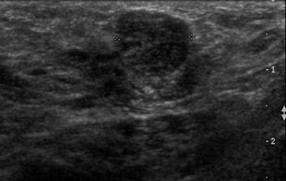
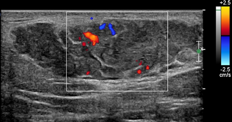
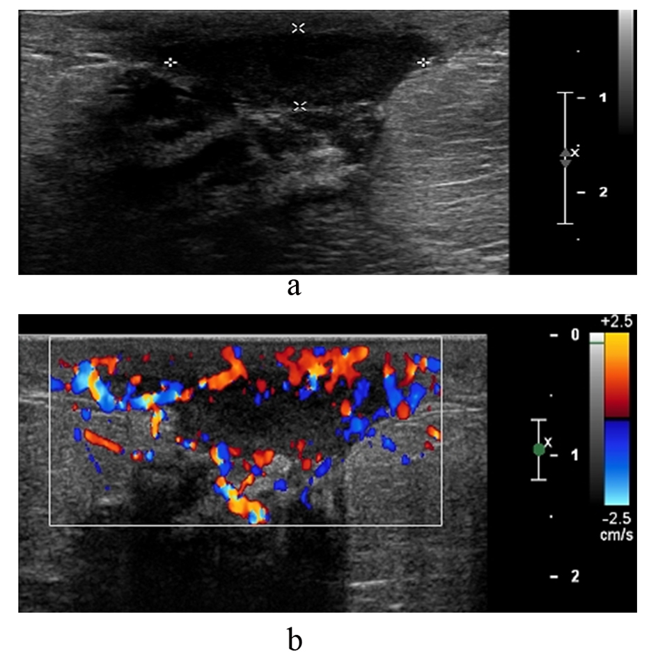
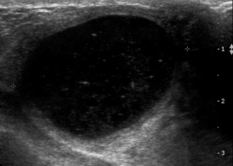
Figure 1. Complex cystic mass on ultrasound. Fine needle aspiration confirmed a galactocele.
Figure 2.
Figure 2. Hypoechoic mass with well circumscribed margins. Confirmed as a fibroadenoma on histopathology.
Figure 3.
Figure 3. Lobulated hypoechoic lesion demonstrating hypervascularity on colour Doppler flow. Histopathology confirmed a lactating adenoma.
Figure 4.
Figure 4. (a and b). This patient was 3 months post-partum and presented with inflammation of the breast. Ultrasound shows a fluid collection in the retroareolar region with hypervascularity of the surrounding tissue. Imaging findings confirmed a retroareolar abscess with mastitis of the adjacent tissue.
Figure 5.
Figure 5. Mass lesion with indistinct margins. The lesion was a grade 2 invasive lobular cancer on histopathology.