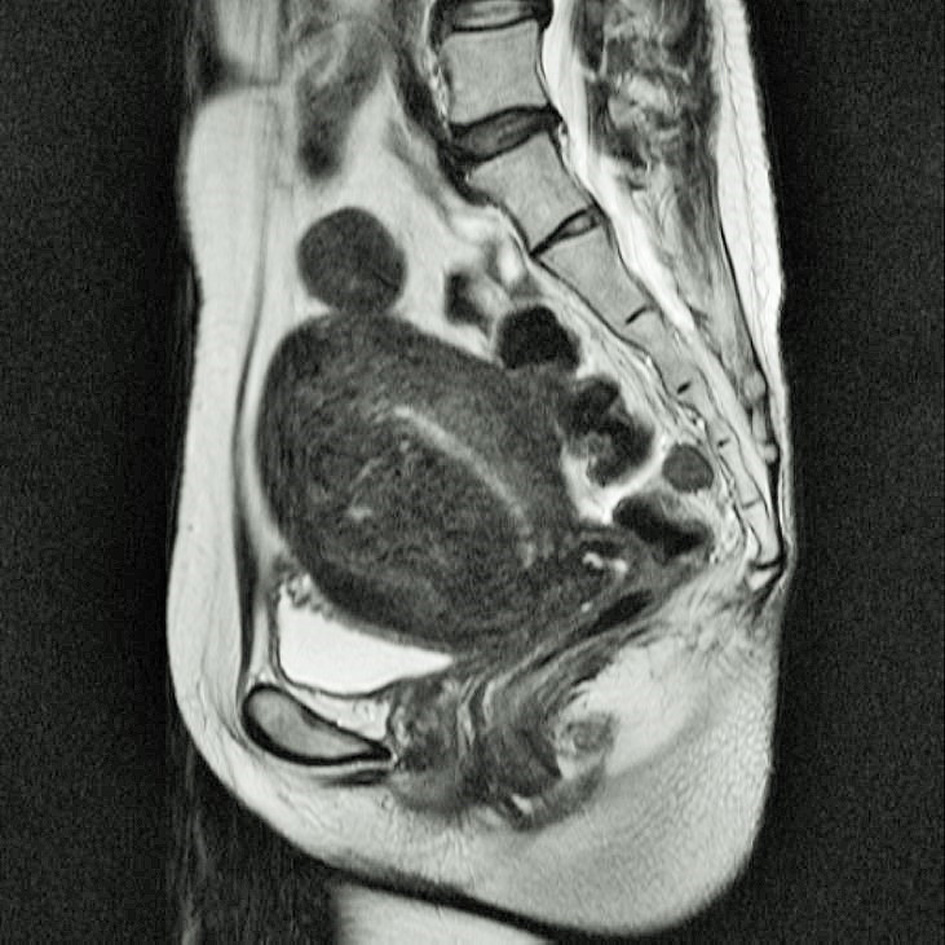
Figure 1. Preoperative MRI of the lower abdomen and pelvis; T2 weighted sagittal section image. Protruded tumor at the uterine fundus 3 cm in diameter and thickening of uterine anterior wall.
| Journal of Clinical Gynecology and Obstetrics, ISSN 1927-1271 print, 1927-128X online, Open Access |
| Article copyright, the authors; Journal compilation copyright, J Clin Gynecol Obstet and Elmer Press Inc |
| Journal website http://www.jcgo.org |
Case Report
Volume 5, Number 1, March 2016, pages 41-44
Delayed Ureteral Insufficiency After Ureteral Obstruction During Total Laparoscopic Hysterectomy: Utility of Cystoscopy for the Intraoperative Detection of Obstruction, and for the Avoidance of Postoperative Surgical Intervention
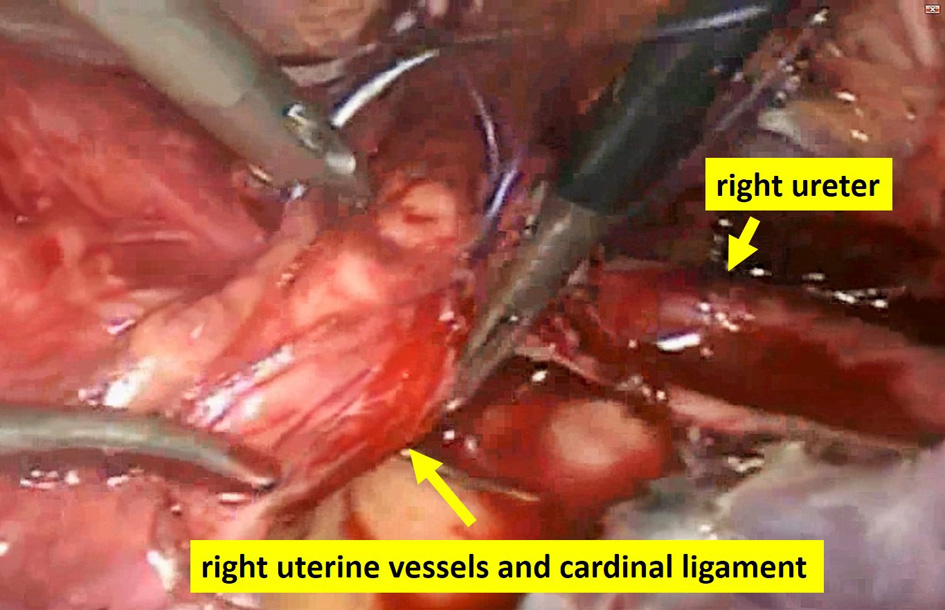
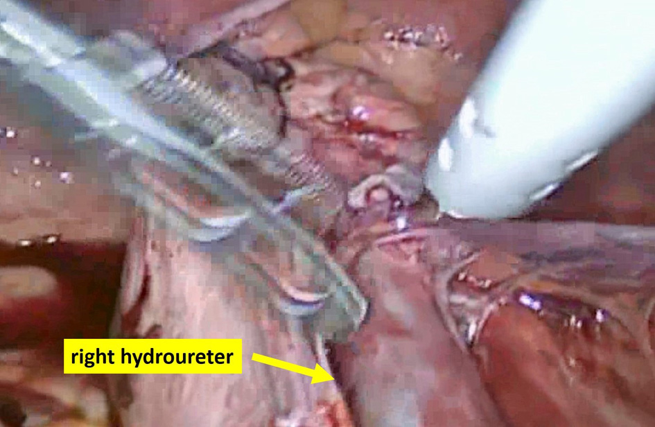
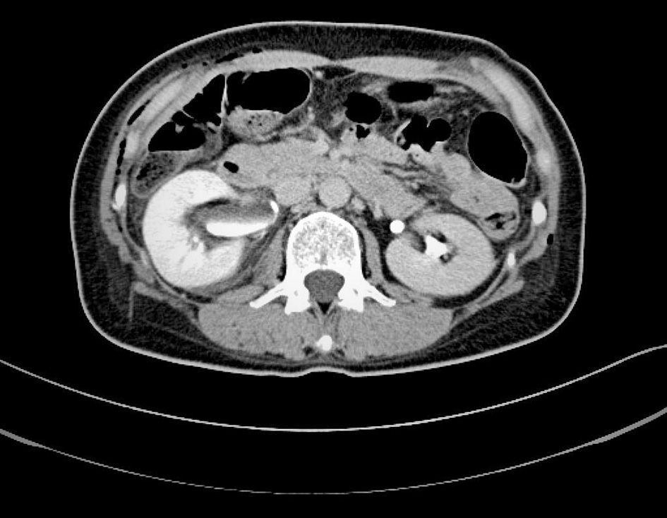
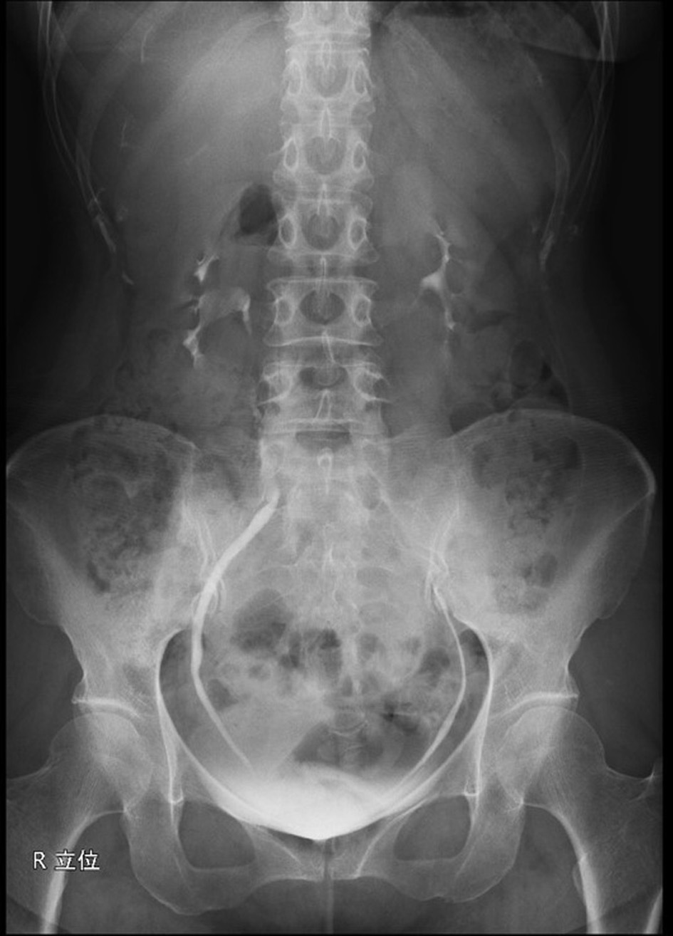
Figures