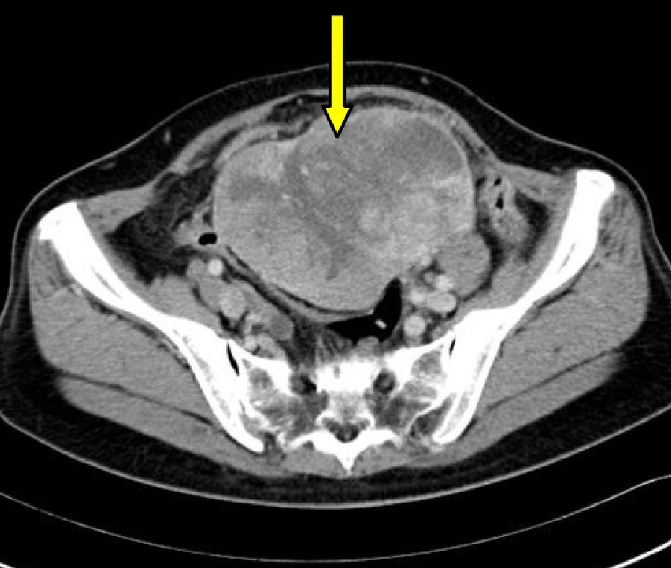
Figure 1. Pelvic computed tomography image: a large mass (arrow) occupying the pelvis, with an irregular shape and contrast enhancement is shown.
| Journal of Clinical Gynecology and Obstetrics, ISSN 1927-1271 print, 1927-128X online, Open Access |
| Article copyright, the authors; Journal compilation copyright, J Clin Gynecol Obstet and Elmer Press Inc |
| Journal website http://www.jcgo.org |
Case Report
Volume 7, Number 1, March 2018, pages 26-29
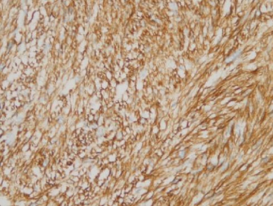
Case Report of a Primary Ovarian Leiomyosarcoma Diagnosed by H-Caldesmon Staining
Figures