Figures
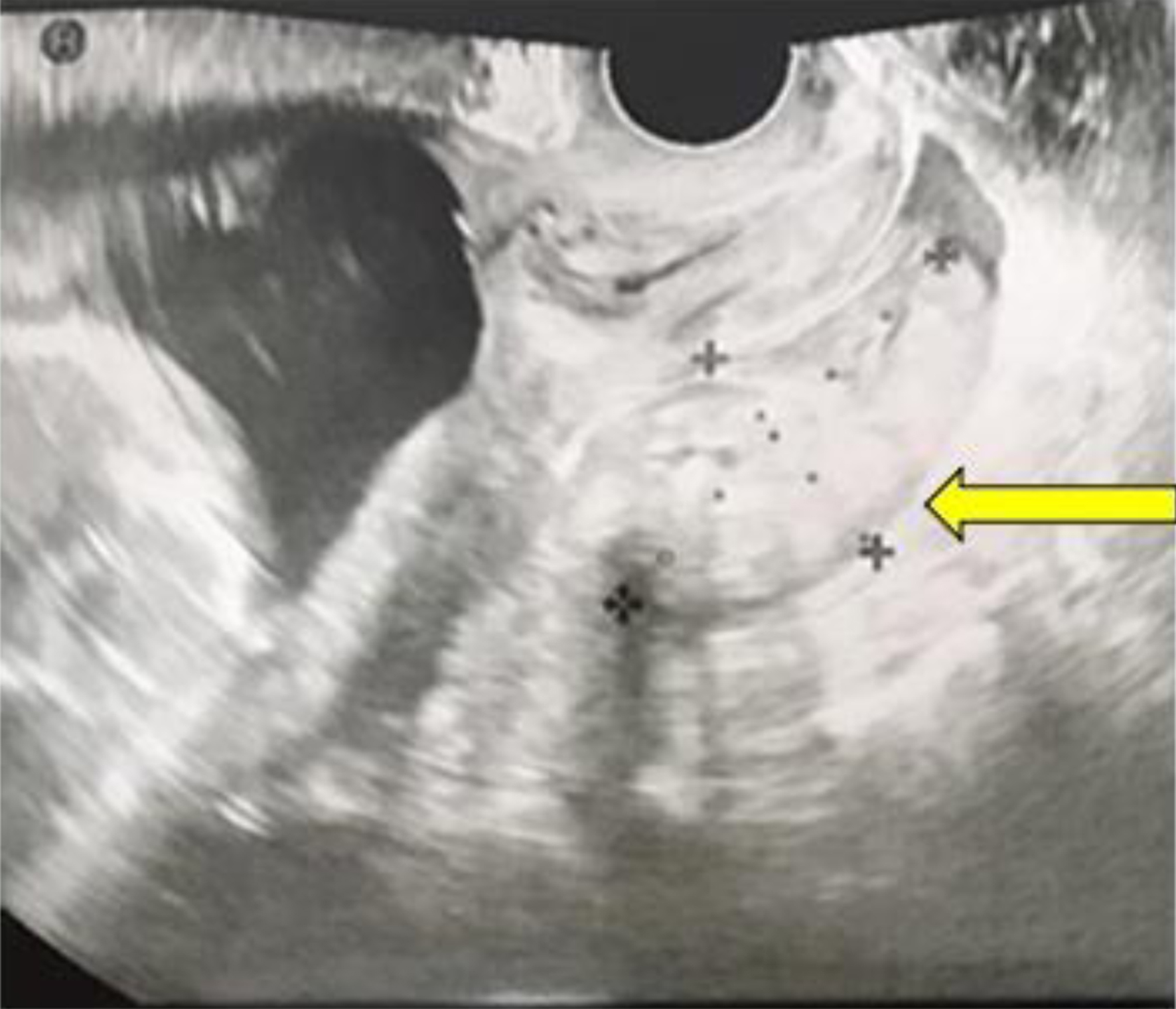
Figure 1. Transvaginal sonography reveals a hyper-echoic lesion measuring 32 × 57 mm located in the Douglas fossa at the time of admission (yellow arrow).
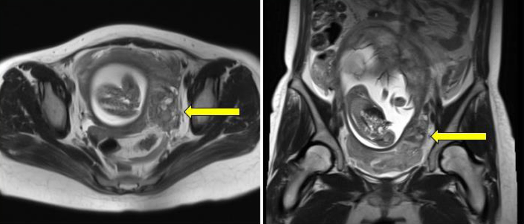
Figure 2. A mass-like lesion is identified in close proximity to the left uterine wall (yellow arrows), exhibiting a heterogeneous signal on both T1- and T2-weighted magnetic resonance images, suggesting a hematoma.
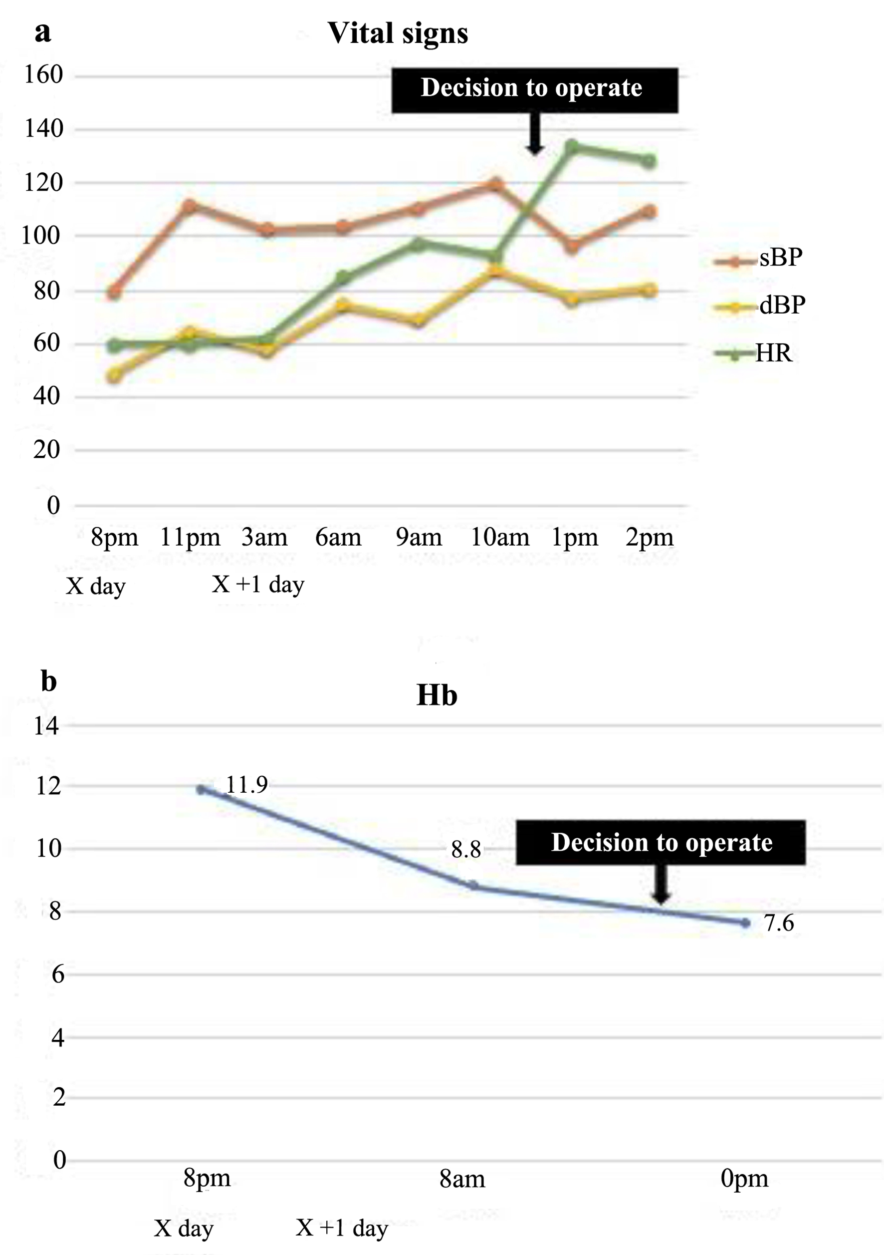
Figure 3. The patient’s vital signs were stable at the time of admission; however, her heart rate increased with time (a), and her hemoglobin levels dropped markedly (b). sBP: systolic blood pressure; dBP: diastolic blood pressure; HR: heart rate; Hb: hemoglobin.
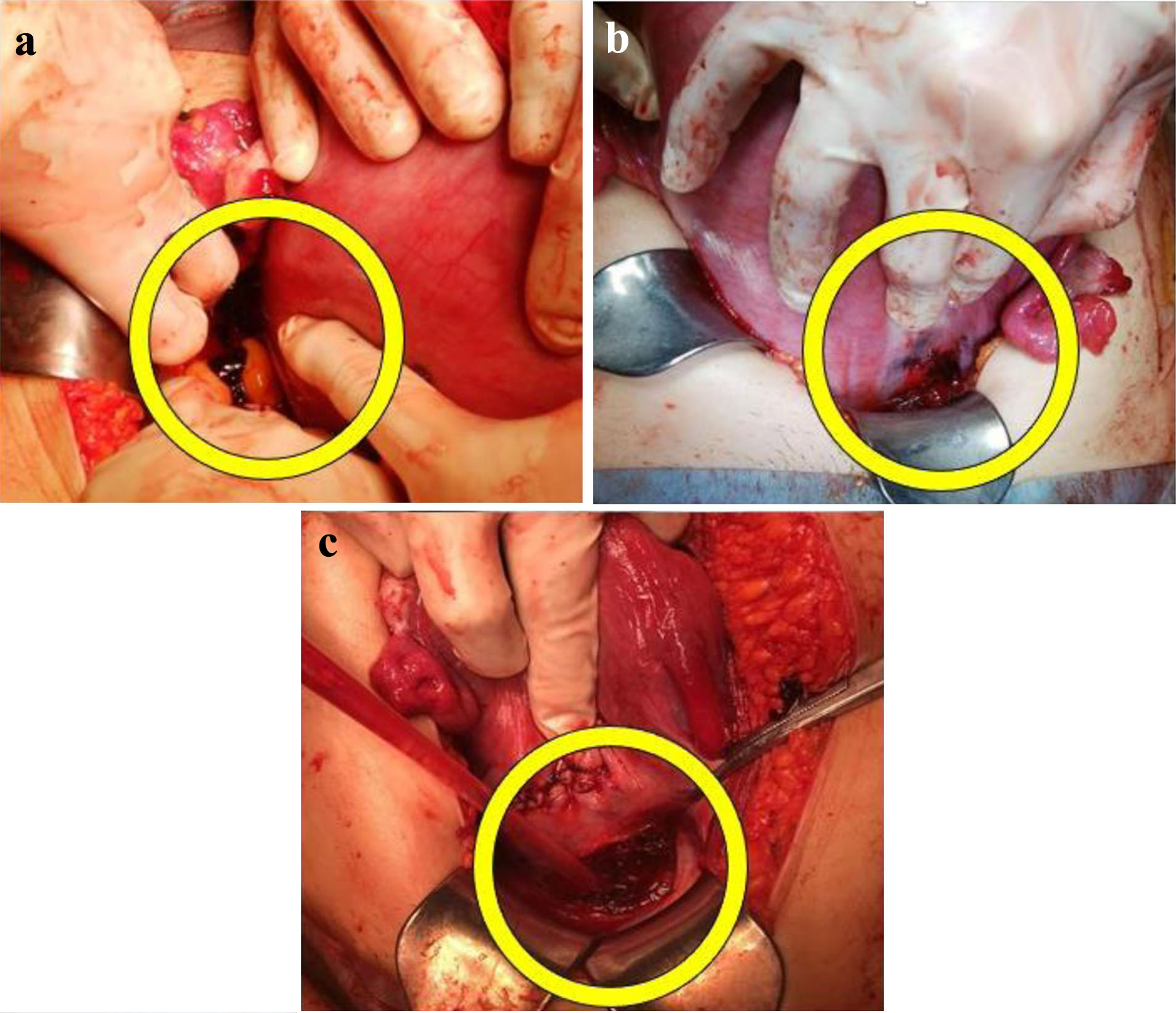
Figure 4. Intraabdominal examination reveals lacerations (yellow circles) on the posterior surface of the lower uterus (a), the anterior surface of the lower uterus (b), and in the vesicouterine pouch peritoneum (c) of the left uterine myometrium.
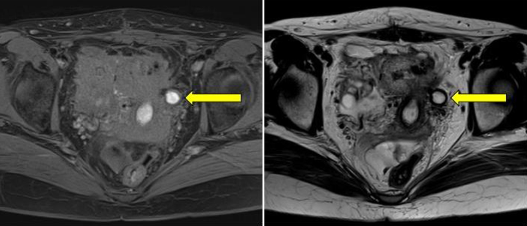
Figure 5. A pelvic magnetic resonance image taken 1 month post-operatively illustrates a cystic lesion (yellow arrows) in close proximity to the left uterine wall, displaying a high signal on both T1- and T2-weighted images, suggesting an endometrial cyst.