Journal of Clinical Gynecology and Obstetrics, ISSN 1927-1271 print, 1927-128X online, Open Access
Article copyright, the authors; Journal compilation copyright, J Clin Gynecol Obstet and Elmer Press Inc
Journal website https://www.jcgo.org

Case Report

Volume 13, Number 2, June 2024, pages 41-47


Condyloma Lata: A Vulvar Manifestation of Secondary Syphilis

Figures

Figure 1.
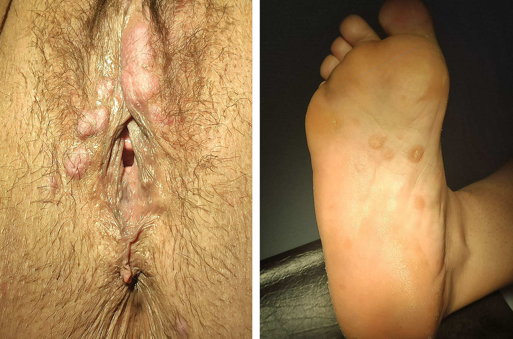
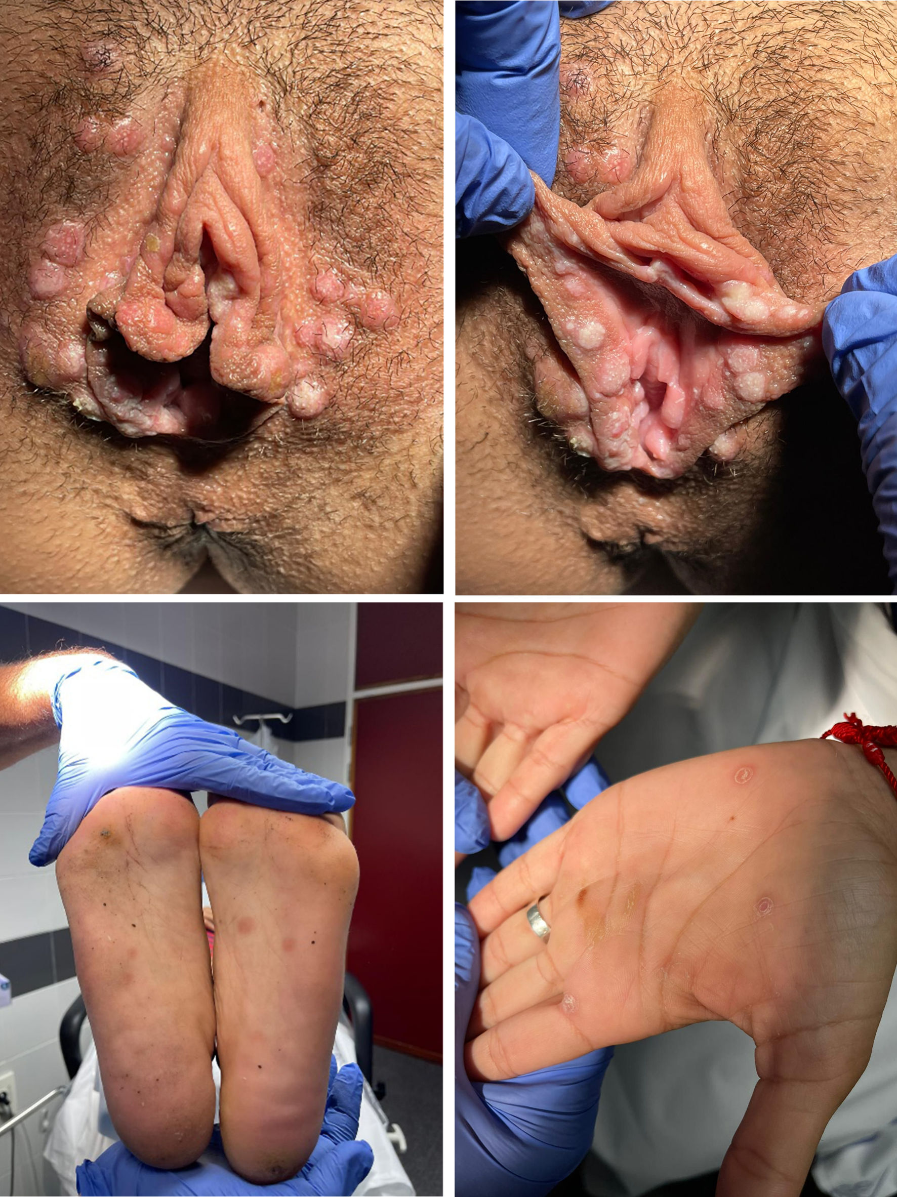
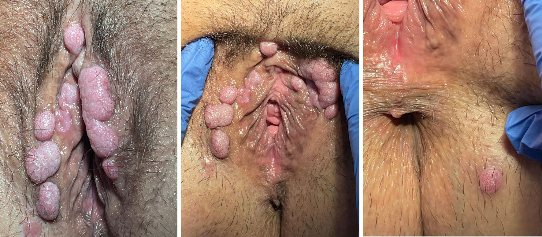
Figure 1. Condyloma lata at presentation (case 1).
Figure 2.
Figure 2. Histopathology of condyloma lata. (a) and Hematoxylin & eosin stain shows intense pseudoepitheliomatous hyperplasia with regular acanthosis at the periphery and a polymorphonuclear infiltrate in the upper layers. (b) Subepithelial examination revealed inflammatory infiltration rich in plasma cells, as well as frequent vasculitis images. (c, d) Treponema immunohistochemistry. The spirochaetes are predominantly arranged in a basal distribution.
Figure 3.
Figure 3. Lesions after treatment with penicillin G benzathine 2.4 million IU (case 1).
Figure 4.
Figure 4. Condyloma lata at presentation (case 2).
Figure 5.
Figure 5. Histopathology of condyloma lata (case 2). (a) Hematoxylin & eosin stain shows epidermal hyperplasia and a dense mixed inflammatory infiltrate with abundant plasma cells. (b) Positive immunohistochemical staining for Treponema was observed.