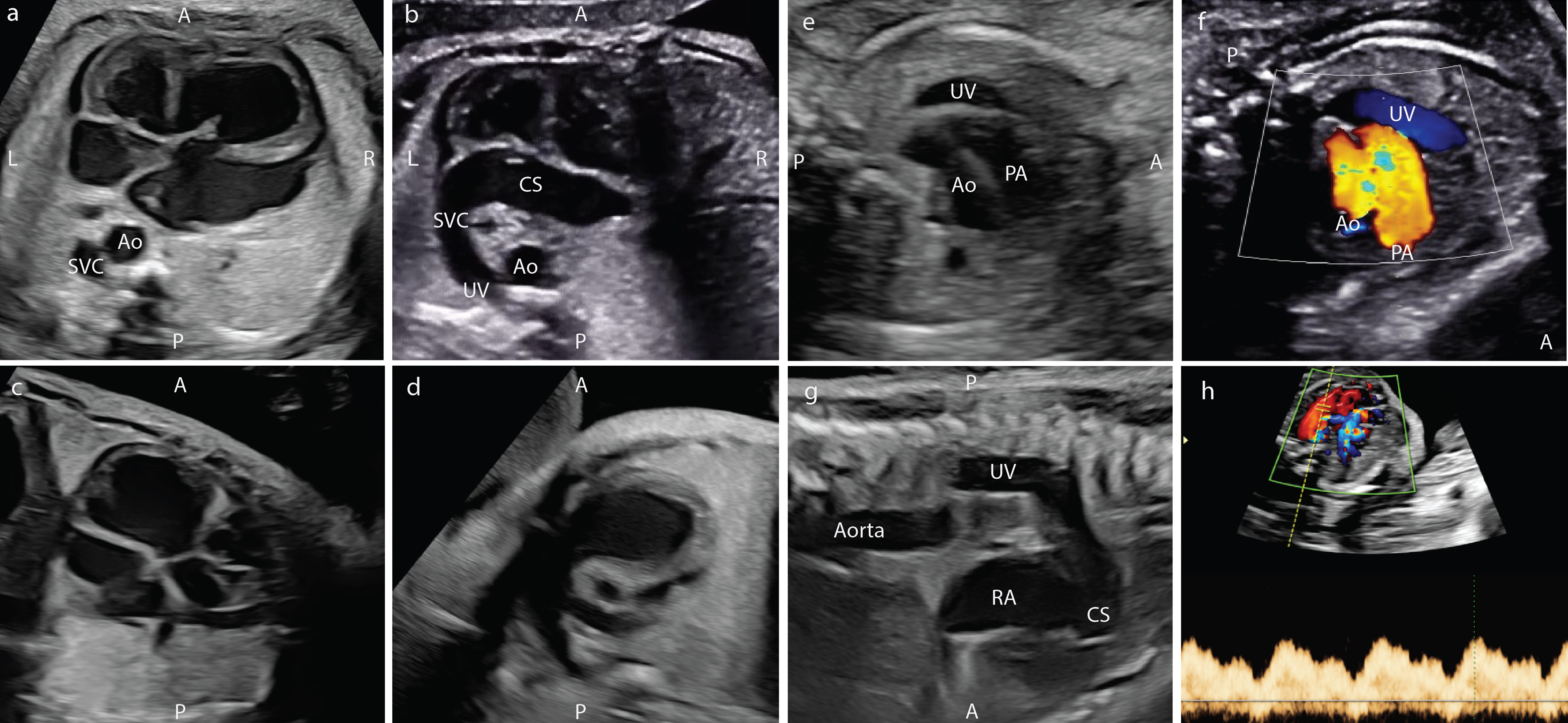
Figure 1. Fetal ultrasound findings in the patient with absent ductus venosus. (a) Four-chamber view with dilated right atrium and ventricle and left SVC. (b) Four-chamber view with dilated coronary sinus connected to left SVC. (c) Left ventricular outflow tract. (d) Right ventricular outflow tract. (e) Three-vessel trachea view with umbilical vein. (f) Three-vessel trachea view with umbilical vein in color Doppler. (g) Sagittal view of left SVC. (h) Doppler of umbilical vein. A: anterior; Ao: aorta; CS: coronary sinus; L: left; LV: left ventricle; PA: pulmonary artery; P: posterior; R: right; RV: right ventricle; SVC: superior vena cava; UV: umbilical vein.