Figure 1. Transvaginal ultrasound image. In the left adnexal area, an echogenic mass with smooth, well-defined borders can be observed (arrows). Posterior acoustic shadowing is also observed.
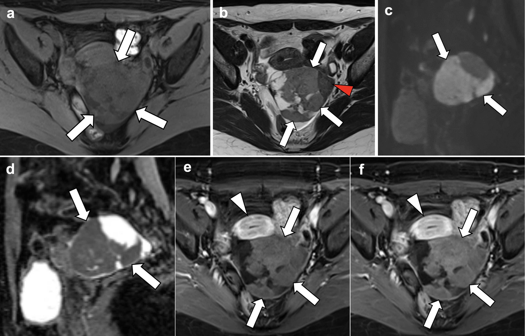
Figure 3. Magnetic resonance imaging. (a) T1-weighted axial section, (b) T2-weighted axial section, (c, d) diffusion-weighted sagittal images, and (e, f) dynamic contrast-enhanced axial images. On T2-weighted imaging, the left ovarian mass is mildly hyperintense in the larger right solid portion (b; arrows), with several cystic areas detected, and hypointense in the smaller left solid portion (arrowhead). On diffusion-weighted imaging, reduced diffusion is observed in the solid portion of the mass (c, d; arrows), which is pathologically proven to be a cellular fibroma component with hypercellularity. On dynamic contrast-enhanced imaging, compared with pre-contrast T1WI, the entire solid portion excluding cystic components of the mass shows a faint and gradual enhancement pattern (e, f; arrows), corresponding to a tumor with abundant fibrous components pathologically proven, whereas the uterine myometrium shows relatively strong enhancement (arrowheads).
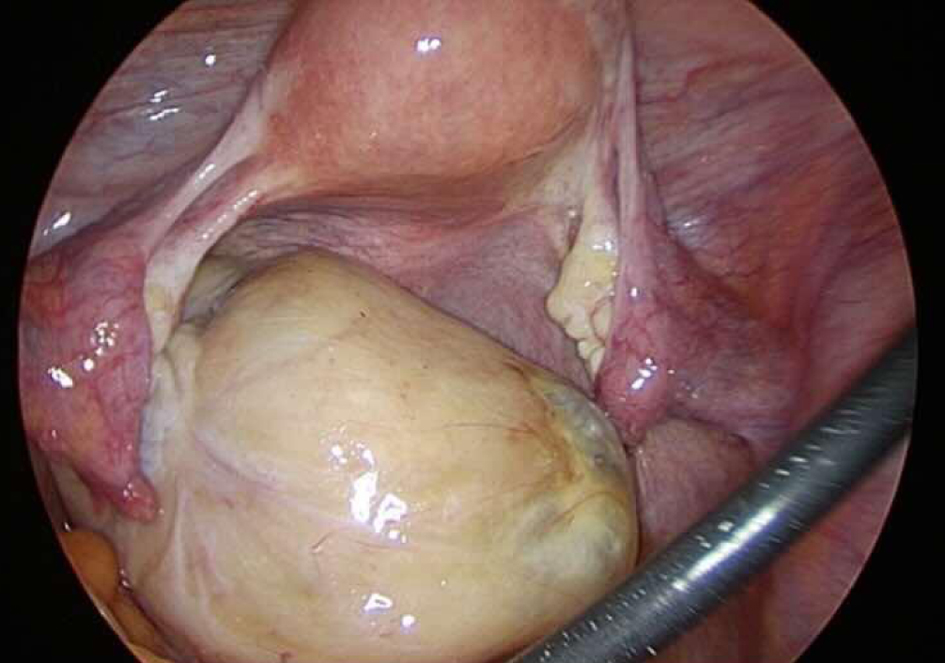
Figure 4. Laparoscopic findings. A yellowish-white solid mass is detected on the left ovary, showing torsion of approximately one rotation without any signs of necrosis. The uterus and right ovary appeared within normal range. No evidence of intraperitoneal dissemination is observed. There is a small amount of ascitic fluid accumulation.
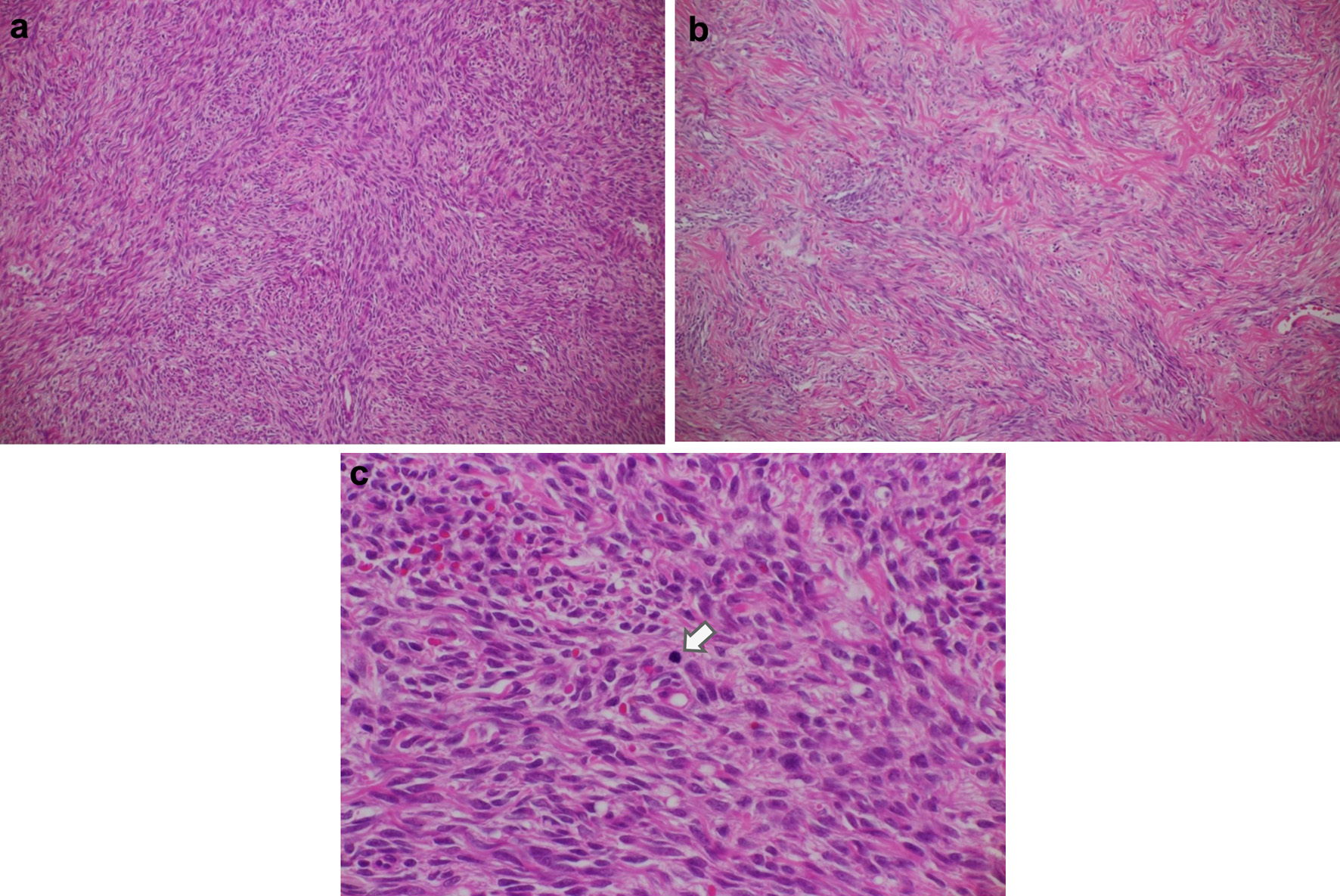
Figure 5. Histologic features of the ovarian tumor. (a) Cellular fibroma (hematoxylin and eosin, original magnification × 100). This pathological specimen shows abundant spindle-shaped cells and fibrocollagenous stroma. Spindle-shaped cells occupy a relatively large area, and mitotic figures are widely found compared with the ordinary fibroma. (b) Ordinary fibroma (hematoxylin and eosin, original magnification × 100). The density of spindle-shaped cells is lower than in (a). (c) Cellular fibroma containing cells undergoing mitosis (arrow) (hematoxylin and eosin, original magnification × 400). Arranged spindle-shaped cells can be seen as well as mitotic figures (average 3.06 mitoses/2.4 mm2). Overall, no prominent findings indicative of extensive necrosis.