Journal of Clinical Gynecology and Obstetrics, ISSN 1927-1271 print, 1927-128X online, Open Access
Article copyright, the authors; Journal compilation copyright, J Clin Gynecol Obstet and Elmer Press Inc
Journal website https://www.jcgo.org

Case Report

Volume 13, Number 3, December 2024, pages 95-100


A Rare Case of Leiomyosarcoma in a Fourteen-Year-Old Female: Challenges in Treatment and Recurrence Management

Figures

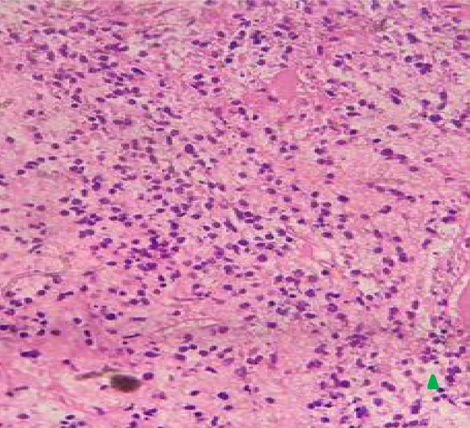
Figure 1.
Figure 1. Histopathology of vaginal uterine mass (× 40) showing endometrial cartilaginous metaplasia upon magnification × 40 (arrow).
Figure 2.
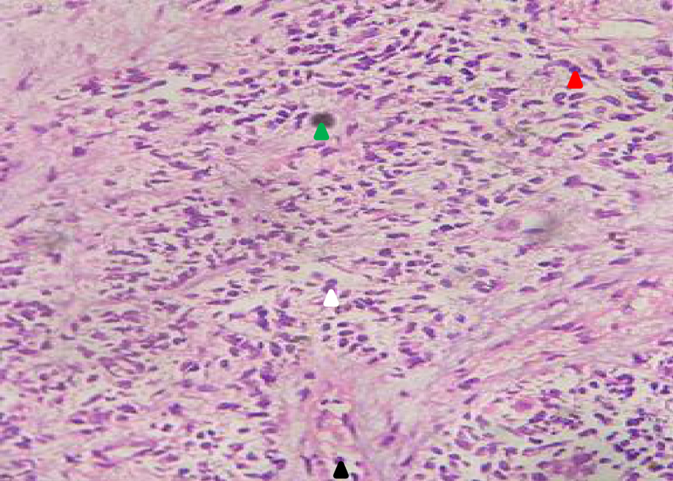
Figure 2. Histopathology of uterine mass (× 40) showing necrotic tissues (green) with rounded, oval, polymorphic nuclear cells (red arrow), hyperchromatic (white arrow), and mitotic tumor mass (black arrow). The presence of endometrial sarcoma was seen in the biopsy result.
Figure 3.
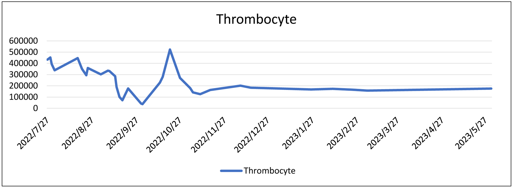
Figure 3. Laboratory trend of thrombocyte level.
Figure 4.
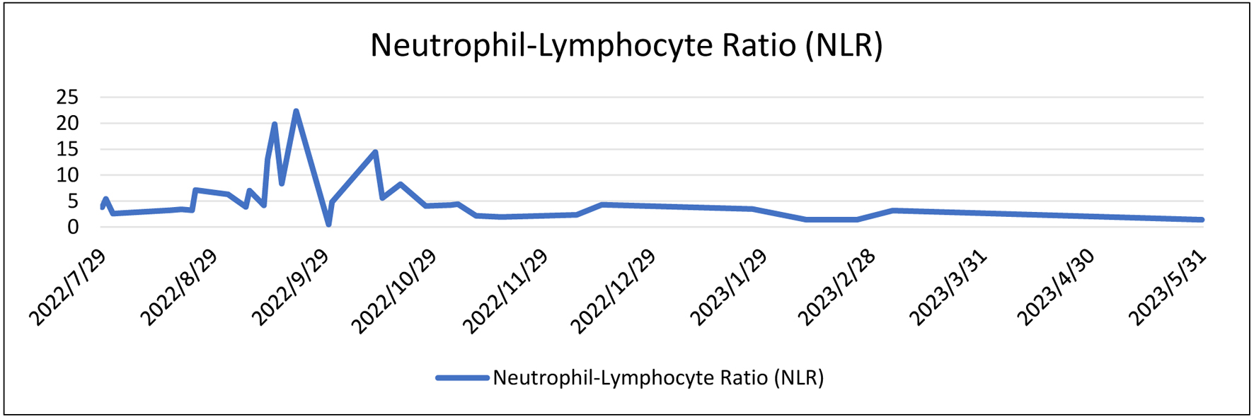
Figure 4. Laboratory trend of neutrophil-lymphocyte level.
Figure 5.
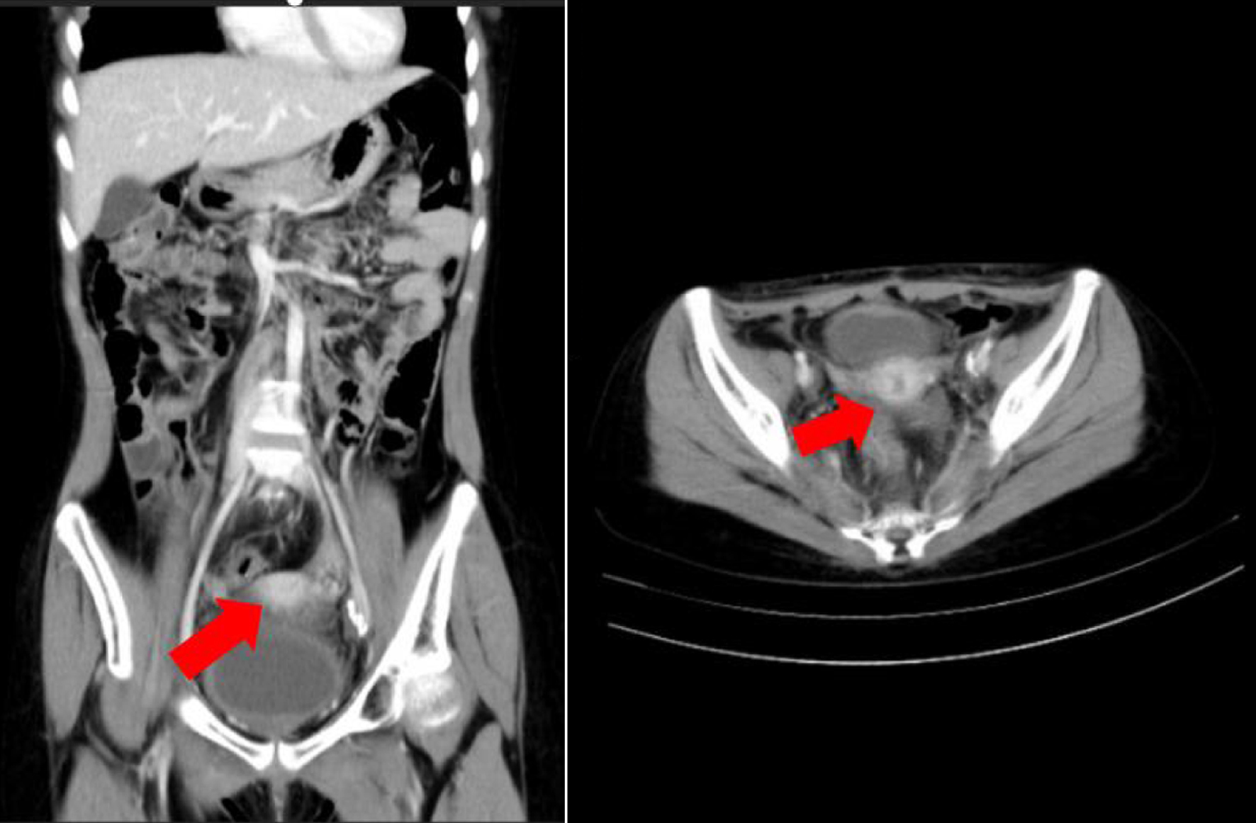
Figure 5. Computed tomography scan of the patient. Red arrow shows lobulated solid mass with calcification at corpus of uterine (dimension 3.05 × 5.13 × 5.05 cm) with fat stranding around, suspected for leiomyosarcoma.