

| Journal of Clinical Gynecology and Obstetrics, ISSN 1927-1271 print, 1927-128X online, Open Access |
| Article copyright, the authors; Journal compilation copyright, J Clin Gynecol Obstet and Elmer Press Inc |
| Journal website http://www.jcgo.org |
Case Report
Volume 8, Number 1, March 2019, pages 29-31
A Case of Ovarian Carcinoid Heart Disease
Tomoko Matsuzonoa, b, Sze Man Wonga, Wai Hon Lia, Yuk May Chana
a2/F, Block F, Queen Elizabeth Hospital, 30 Gascoigne Road, Hong Kong, China
bCorresponding Author: Tomoko Matsuzono, 2/F, Block F, Queen Elizabeth Hospital, 30 Gascoigne Road, Hong Kong, China
Manuscript submitted March 3, 2019, accepted March 12, 2019
Short title: Ovarian Carcinoid Heart Disease
doi: https://doi.org/10.14740/jcgo539
| Abstract | ▴Top |
We presented a 68-year-old lady who had total abdominal hysterectomy and bilateral salpingo-oophorectomy performed for a 7 cm complex ovarian tumor and ascites on ultrasound scan. Histopathology report confirmed the diagnosis of ovarian carcinoid tumor. This lady presented with gross ascites post-operatively, echocardiogram revealed right heart overload, tricuspid and pulmonary valve regurgitation compatible with carcinoid heart disease. Tricuspid and pulmonary heart replacement was performed for this lady and she had an uneventful recovery.
Keywords: Carcinoid syndrome; Carcinoid heart disease; Hysterectomy
| Introduction | ▴Top |
Neuroendocrine tumors are a heterogeneous group of neoplasms most commonly occurring in the gastrointestinal tract or the lungs, although they may arise less frequently in other sites [1]. Primary ovarian carcinoid tumors constitute only about 1% of all carcinoid tumors and < 0.1% of all ovarian carcinomas. Carcinoid syndrome is characterized by diarrhea, vasomotor changes, bronchospasm and symptoms of carcinoid heart disease, which affects approximately one-third of the patients with carcinoid tumors. Carcinoid syndrome is due to serotonin-like substances released directly into the systemic circulation through the ovarian venous system. Carcinoid heart disease occurs in one-third to half of the patients with carcinoid syndrome, with tricuspid or pulmonary regurgitation being the most common valves affected [2]. However, patients with carcinoid heart disease can be relatively asymptomatic; therefore, diagnosis can be difficult until late presentation of heart failure.
| Case Report | ▴Top |
We presented a 68-year-old lady who was incidentally noted to have a 7 cm ovarian mass with ascites on an ultrasound scan for the workup for her thrombocytopenia.
On ultrasound scan, she had a thickened endometrial lining of up to 8.3 mm, there was a right complex mixed echogenic adnexal mass and small amount of ascitic fluid. Computed tomography (CT) scan of the abdomen and pelvis confirmed a 7 cm lobulated enhancing mass over the right adnexal region, suspected right ovarian in origin, moderate amount of ascites, and no obvious lymphadenopathy. Pre-operative cancer antigen 125 (CA125) was 17U/mL. Total abdominal hysterectomy and bilateral salpingo-oophorectomy was performed, post-operative course was unremarkable and she was discharged on day 9 post-operatively. Final histopathology report confirmed teratoma with carcinoid tumor and no evidence of malignancy, and hence, no adjuvant treatment was required for this lady.
However, this lady was readmitted 1 month later for abdominal distension. CT scan revealed gross ascites, and mild peritoneal thickening of unknown significance. Repeated paracentesis was negative for any infective component (negative for Gram stain, acid fast bacilli smear, culture and sensitivity), malignancy, and chyle. Peritoneal fluid for creatinine was normal at 47 µmol/L, and blood test revealed a normal liver and renal function as well as a normal albumin level. In view of negative findings of apparent cause of ascites, the patient was put on conservative management with interval reassessment scan.
Reassessment CT scan 2 months later identified persistent ascites, engorged hepatic veins, inferior vena cava, and right atrium (Fig. 1). Echocardiogram revealed right heart overload with tricuspid and pulmonary valve regurgitation. Given the history of ovarian carcinoid tumor, the diagnosis of carcinoid heart disease was made.
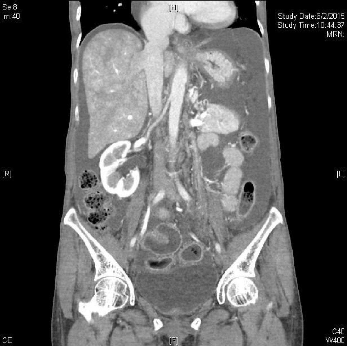 Click for large image | Figure 1. Post-operative CT scan showing gross ascites, engorged hepatic veins and inferior vena cava. |
The patient finally underwent a tricuspid and pulmonary heart replacement and recovered uneventfully.
| Discussion | ▴Top |
Neuroendocrine tumors are a heterogeneous group of neoplasms most commonly occurring in the gastrointestinal tract or the lungs, although they may arise less frequently in other sites [1]. Primary ovarian carcinoid tumors constitute only about 1% of all carcinoid tumors and < 0.1% of all ovarian carcinomas. The histologic subtypes can be divided into four groups: insular, trabecular, strumal (struma ovarii and carcinoid), and mucinous with or without an associated mature teratoma. [2]. Carcinoid syndrome is characterized by facial flushing, bronchospasm, edema and diarrhea, and has been estimated to occur in one-third of patients with ovarian carcinoid tumors. The syndrome manifestation was due to serotonin-like substances released directly into systemic circulation through the ovarian venous system bypassing hepatic deactivation. In contrast, gastrointestinal carcinoids lack direct access to the vena cava and are reported to cause carcinoid syndrome infrequently, unless there is significant metastatic liver involvement [3].
Carcinoid heart disease, which represents the development of plaque-like, fibrous endocardial thickening involving heart valves, mainly in the right heart, is a frequent occurrence in patients with carcinoid syndrome. In patients with carcinoid heart disease, right atrial and right ventricular enlargement is present in up to 90% of cases; and ventricular septal wall motion abnormalities are seen in almost half of the cases. The tricuspid valve leaflets and subvalvular structures are often thickened, shortened and retracted, leading to incomplete coaptation and usually moderate or severe tricuspid regurgitation [4].
Carcinoid heart disease of different grades of severity has been reported in 40-50% of patients with carcinoid syndrome [5]. The main predictor of clinical outcome in patients with carcinoid syndrome is the extent and severity of cardiac involvement, therefore, transesophageal echocardiography (TTE) plays a central role in the diagnostic and prognostic evaluation of this condition. It is recommended to be performed in all patients with carcinoid syndrome with suspicion of carcinoid heart disease.
Carcinoid heart disease is a major source of morbidity and mortality for patients with carcinoid syndrome. It typically leads to progressive dysfunction of the valves involved (mostly tricuspid and pulmonary) and disability of the patient. Without treatment, the prognosis of carcinoid heart disease is poor, with 3-year survival as low as 31% [5].
Although carcinoid tumors are rare malignancies, cardiac involvement is relatively common. Despite severe disease, patients may possess relatively few signs or symptoms in the early stages. Echocardiography is the gold standard of choice of investigation which reveals a unique valvular appearance. Increasing severity of valvular disease can lead to right heart failure and major morbidity and mortality of patients. A high awareness of the possibility of carcinoid heart disease in patient with carcinoid tumor, and a multidisciplinary team approach is required to provide informed decisions on the optimal management for this group of complex patients.
Acknowledgments
This study was supported by the Obstetrics and Gynecological Department of Queen Elizabeth Hospital.
Financial Disclosure or Funding
None to declare.
Conflict of Interest
Authors declare there is no conflict of interest.
Informed Consent
Not applicable.
Author Contributions
Authors are involved in the multidisciplinary care of this lady.
| References | ▴Top |
- Reed NS, Gomez-Garcia E, Gallardo-Rincon D, Barrette B, Baumann K, Friedlander M, Kichenadasse G, et al. Gynecologic Cancer InterGroup (GCIG) consensus review for carcinoid tumors of the ovary. Int J Gynecol Cancer. 2014;24(9 Suppl 3):S35-41.
doi pubmed - Davis KP, Hartmann LK, Keeney GL, Shapiro H. Primary ovarian carcinoid tumors. Gynecol Oncol. 1996;61(2):259-265.
doi pubmed - Motoyama T, Katayama Y, Watanabe H, Okazaki E, Shibuya H. Functioning ovarian carcinoids induce severe constipation. Cancer. 1992;70(2):513-518.
doi - Fox DJ, Khattar RS. Carcinoid heart disease: presentation, diagnosis, and management. Heart. 2004;90(10):1224-1228.
doi pubmed - Davar J, Connolly HM, Caplin ME, Pavel M, Zacks J, Bhattacharyya S, Cuthbertson DJ, et al. Diagnosing and managing carcinoid heart disease in patients with neuroendocrine tumors: an expert statement. J Am Coll Cardiol. 2017;69(10):1288-1304.
doi pubmed
This article is distributed under the terms of the Creative Commons Attribution Non-Commercial 4.0 International License, which permits unrestricted non-commercial use, distribution, and reproduction in any medium, provided the original work is properly cited.
Journal of Clinical Gynecology and Obstetrics is published by Elmer Press Inc.